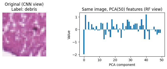
Spatial filters captured the local texture signal.
PathMNIST Histology Classification
A comparative study of Random Forest, multilayer perceptron, and convolutional neural network models on nine colorectal tissue patch classes. The result shows how much spatial structure matters for low resolution histology images.
Local PathMNIST samples exported from the included training split
Measured on the held out 8,000 image test set.
Class balanced score across all nine categories.
Model Comparison
Final models were trained from the selected hyperparameters and evaluated on the same test split.
Accuracy and Macro F1
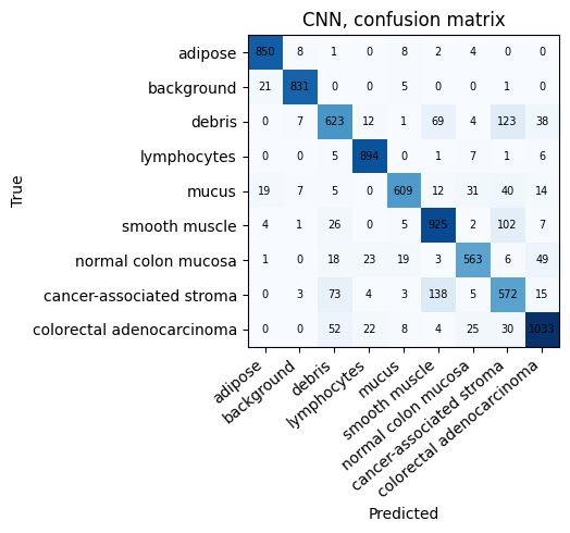
CNN creates the clear separation.
Training Cost
Accuracy came with a longer CPU run.
Interactive Results
Switch between confusion matrices, tuning searches, and training histories without losing the comparison context.
Class Explorer
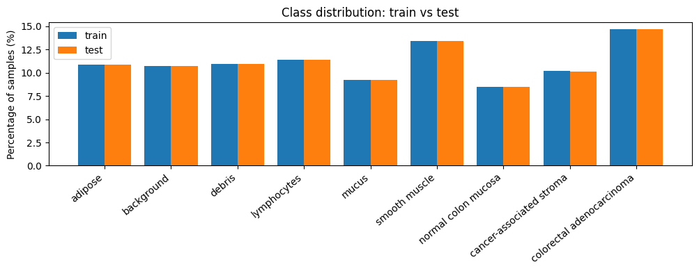
Review sample patches, class balance, and per class F1 scores for each tissue category.
Method Notes
The modelling choices are kept visible so the result page remains audit friendly for a technical reader.
Dataset
The assignment subset contains 32,000 training images and 8,000 test images. Every patch is a 28 by 28 RGB crop from PathMNIST.
Preprocessing
Random Forest used flattened pixels with PCA, MLP used flattened standardized vectors, and CNN kept the image tensor shape.
Interpretation
Flat pixel models struggled on glandular and stromal texture. The CNN improved the clinically important normal mucosa and adenocarcinoma classes, but this is coursework and not a diagnostic system.